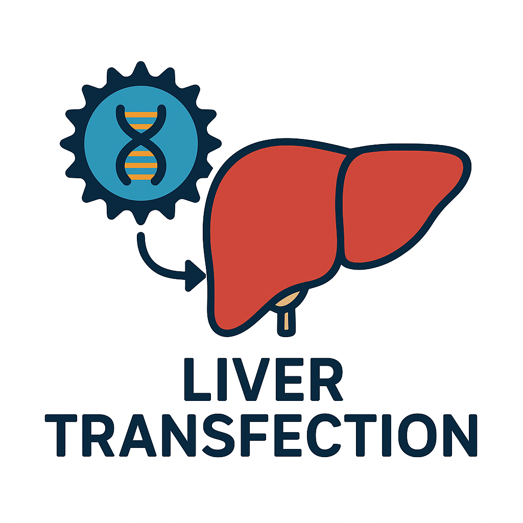
Intracellular Trafficking and Nuclear Import of Transfected Plasmid DNA in Hepatocytes
Once foreign DNA is introduced into a hepatocyte (via lipofection, electroporation, viral vectors, etc.), its ultimate goal is to enter the nucleus where transcription occurs. The path from the cell membrane to the nucleus is fraught with challenges. Endocytic vesicles can trap the DNA and shunt it to lysosomes for degradation; the crowded cytoplasm and cytoskeletal network must be navigated; and finally, the nuclear envelope must be crossed. This section focuses on intracellular trafficking and nuclear import of transfected plasmid DNA in liver cells, dissecting each stage of the journey.
We first outline the general barriers: endosomal escape and cytosolic transport, then emphasize the nuclear import step, which is often the rate-limiting barrier for gene delivery in non-dividing cells. Hepatocytes in adult liver are largely quiescent (non-dividing), meaning their nuclear envelopes remain intact; unlike in dividing cells, plasmids cannot simply “wait” for mitosis when the nuclear membrane breaks down. We will examine how some plasmid sequences and cellular mechanisms assist DNA in threading through nuclear pore complexes. We also consider how transfection reagents can be engineered to improve nuclear delivery – for example, by attaching nuclear localization signals (NLS) or utilizing the cell’s importin transport system.
Endosomal Uptake and Cytosolic Transport
Most non-viral transfection vectors introduce DNA into hepatocytes via endocytosis. Cationic liposomes and polymers form complexes with DNA that bind to the cell membrane and get internalized in endosomal vesicles. Once inside endosomes, the DNA must escape into the cytosol before the endosome matures into a lysosome (where DNases like DNase II would destroy it. Many transfection reagents are designed with this challenge in mind. Polyethyleneimine (PEI), for instance, is thought to facilitate endosomal escape by the “proton sponge” effect – as endosomes acidify, PEI buffers the pH, causing osmotic swelling and vesicle rupture, releasing DNA into the cytosol. Lipid nanoparticles often include helper lipids (like DOPE, a fusogenic lipid) that promote endosomal membrane fusion and disruption.
Following endosomal escape, plasmid DNA finds itself in the cytosolic environment. Here, the DNA is vulnerable to nucleases and may also trigger innate immune sensors (as discussed in the previous section on inflammation). The cell has cytosolic 3’ exonucleases such as TREX1 that readily degrade free DNA to maintain homeostasis. Thus, there is a time pressure for plasmids: they need to reach the nucleus relatively quickly before being degraded. The diffusion of large DNA (~5 kb plasmid is ~10^6 daltons) in the cytoplasm is slow and impeded by the viscoelastic cytosol. However, cells have active transport mechanisms along the cytoskeleton that viruses and some synthetic vectors can hijack. Microtubules radiating from the periphery to the nucleus provide “highways” for cargo. There is evidence that DNA complexes can bind molecular motors; for example, certain peptides on DNA nanoparticles can engage dynein motors for retrograde transport along microtubules. Virus-mimicking strategies have attempted to decorate DNA polyplexes with motor-binding peptides to enhance movement toward the nucleus. In hepatocytes, which are polarized cells, movement to the nucleus may also be influenced by their geometry and the localization of microtubule organizing centers.
Notably, research has shown that simply reaching the perinuclear cytoplasm is not enough – the nuclear membrane remains a formidable gatekeeper. This became clear from classical experiments where microinjection was used: Capecchi’s 1980 study demonstrated that plasmids injected into the nucleus express efficiently in nearly all cells, but the same plasmids injected into the cytoplasm yielded virtually no expression. The difference was stark: a direct nuclear delivery had 50–100% of cells expressing, whereas cytoplasmic delivery of thousands of plasmid copies per cell resulted in none expressing. This underscores that nuclear entry is the critical step that plasmids struggle with inside cells.
Nuclear Import of Plasmid DNA
The nuclear envelope has nuclear pore complexes (NPCs) that allow regulated exchange of material. Small molecules and proteins under ~40 kDa can diffuse through NPCs, but a supercoiled plasmid (several MDa in size) is far beyond that limit. Non-dividing cells thus require an active mechanism for large DNA to enter – likely through the nuclear import machinery that usually ferries proteins. Plasmids do not have proteins of their own, but they can associate with proteins in the cytosol. Some studies suggest that certain DNA sequences can recruit import proteins. One landmark finding was that the SV40 virus origin/enhancer sequence on a plasmid dramatically improved its nuclear import in non-dividing cells. Dean and colleagues (1999) showed that a 366 bp fragment containing the SV40 origin of replication and enhancer was required for efficient nuclear entry of a plasmid in non-dividing cultured cells. They pinpointed the tandem 72-bp repeats of the SV40 enhancer as key – plasmids with that sequence could transit into the nucleus and express at low copy number, whereas those without it remained excluded until cell division occurred. The SV40 enhancer likely works by binding host transcription factors (like AP-1 or TEAD proteins) or replication proteins (like SV40 T-antigen, if present) that themselves carry nuclear localization signals, effectively piggybacking the DNA into the nucleus. By contrast, other strong promoters like the CMV promoter or RSV LTR did not confer import ability. Thus, nuclear import of plasmid DNA is sequence-specific, not automatic for all DNA.
How do these DNA-protein complexes traverse the NPC? One model is that DNA bound by certain proteins (e.g., transcription factors with NLS sequences) can be recognized by the importin-α/β system – the same system that imports NLS-bearing proteins. Importin-α binds the NLS on a protein and importin-β mediates docking and transit through the pore. If a plasmid is coated with histones or other proteins in the cytosol, these could provide the necessary signals. Indeed, plasmid DNA in cells often associates with histones and forms a “minichromatin.” Some transfection formulations even include histone peptides or nuclear targeting peptides to exploit this pathway. For example, attaching multiple NLS peptides to a polylysine-DNA conjugate has been shown to increase nuclear uptake in some cell types. In hepatocytes, these approaches have not been extensively clinically tested, but conceptually, giving the plasmid an NLS tag via binding proteins can improve nuclear import.
It is worth noting that cell cycle effects are pivotal. In dividing cell cultures of liver-derived cell lines (like HepG2 hepatoma cells), transfection is more efficient because during mitosis the nuclear envelope disassembles, allowing plasmids to access chromatin directly. However, primary hepatocytes or quiescent cell lines require plasmids to find their way through NPCs. Some success has been found with electroporation, which can deliver DNA directly into the nucleus if timed with the cell cycle, or by using high-voltage pulses that momentarily permeabilize the nuclear membrane.
Altogen Biosystems addresses some of these challenges through the design of their transfection kits for liver cells. For instance, the Altogen HepG2 Transfection Kit includes a “complex condenser” and “transfection enhancer”altogen.com, which likely help compact the DNA and facilitate its uptake and possibly trafficking. The HepG2 reagent boasts high efficiency and even the ability to generate stable cell lines (indicating DNA reaching the nucleus and integrating or replicating). The consistency of transfection with Altogen’s reagent suggests that it reliably moves DNA into the vicinity of the nucleus in a high percentage of cells. While the proprietary formulation isn’t fully disclosed, such kits often use combinations of lipids and polymers that ensure endosomal escape and perhaps some nuclear localization aid. The result is that one can routinely get >90% transfection of HepG2 cells for siRNA delivery and high plasmid DNA delivery efficiency as well. Those plasmids still face the nuclear import issue, but if HepG2 cells are dividing, many plasmids will get in during mitosis. In non-dividing primary hepatocytes, using similar reagents, one might rely on high plasmid dosage and any nuclear-targeting elements on the plasmid to maximize chances of nuclear entry.
Another aspect of nuclear import is integration vs. episomal persistence. If plasmids manage to integrate into the host genome (either randomly or via transposon systems), then they effectively bypass the long-term nuclear import problem – once integrated, they replicate with the host DNA and are present after cell division. However, random integration frequency for plasmids in the liver is extremely low. Researchers often incorporate systems like Sleeping Beauty or PiggyBac transposons to achieve integration in hepatocytes for stable expression. These systems entail co-delivery of a transposase along with the gene of interest (the transposon). The transposase can cut and paste the transposon into the host DNA. The transposase enzyme, being a protein, has its own NLS and enters the nucleus, carrying out integration. Thus transposon-based vectors cleverly delegate the nuclear entry problem to the transposase protein, rather than the plasmid itself – the plasmid only needs to be in the cytoplasm for transposase to act on it. This highlights an innovative way to overcome nuclear import limitations for non-viral delivery.
Conclusion
The efficiency of liver cell transfection is profoundly influenced by what happens after the DNA is inside the cell. From escaping endosomes to traversing the cytosol and penetrating the nuclear barrier, each step can bottleneck the process. The nuclear import step stands out as a particularly stringent barrier: plasmid DNA in non-dividing hepatocytes struggles to cross the nuclear envelope, which is why gene delivery is often inefficient in vivo. Nonetheless, research has identified solutions: certain DNA elements (like the SV40 enhancer) markedly improve nuclear uptake, and cell-cycle-independent import can be aided by leveraging the cell’s own import machinery through NLS signals.
Innovative transfection technologies and reagents incorporate these insights. The use of targeting sequences, motor protein engagement, and advanced carriers (like Altogen’s liver-specific formulations) helps shepherd plasmid DNA closer to the nucleus and promote its entry. For example, Altogen’s transfection kits for hepatocyte cell lines achieve high efficiency, implying that they effectively surmount early barriers so that the majority of cells receive DNA and have a chance to express it. As we improve delivery to the nucleus – whether by smarter vector design or by transiently opening nuclear pores – the result will be higher expression levels and more reliable outcomes in liver transfection experiments. Understanding these intracellular logistics is therefore key to advancing both gene therapy and basic research involving liver cells.
In summary, the plasmid’s path to the hepatocyte nucleus is complex but increasingly navigable with modern techniques. Each barrier can be addressed: endosomal escape via fusogenic carriers, cytosolic transport via size reduction or motor assistance, and nuclear import via nuclear-targeted sequences or exploitation of cell division. By integrating these strategies, we can achieve efficient nuclear delivery of therapeutic genes to the liver with non-viral methods, complementing viral vectors and expanding the toolkit for treating liver diseases and modeling them in experimental systems.
Sources: Nuclear import barrier description; Capecchi microinjection result; SV40 enhancer effect on plasmid import; Altogen HepG2 transfection kit features.