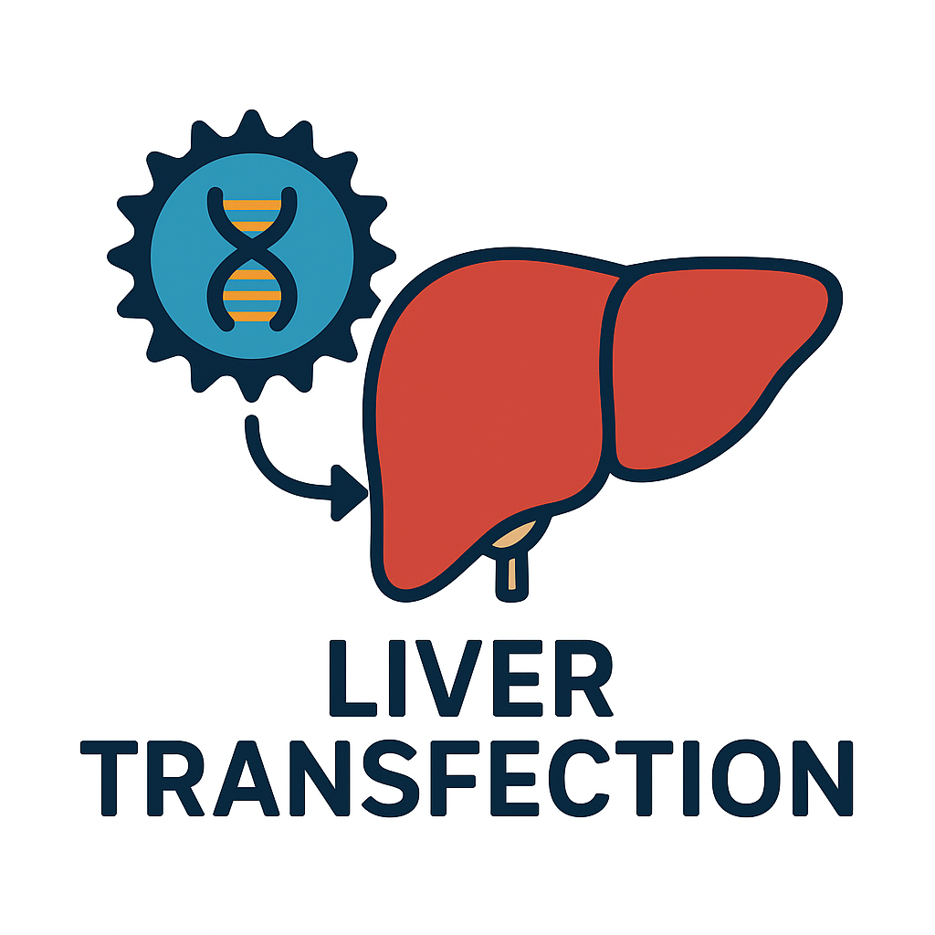
Mechanisms of Endosomal Escape in Lipid Nanoparticle-Mediated Liver Transfection
Lipid nanoparticles (LNPs) have revolutionized the delivery of nucleic acids to the liver, enabling therapies like siRNA drugs and mRNA vaccines. A critical step in LNP-mediated transfection is endosomal escape – the process by which the payload (DNA or RNA) is released from endosomal vesicles into the cytosol after cellular uptake. Endosomal escape is often the efficiency bottleneck: although hepatocytes readily endocytose LNPs, only a small fraction of internalized nucleic acid typically avoids lysosomal degradation and reaches the cytoplasm where it can function. This section explores the mechanisms by which LNPs achieve endosomal escape, including the role of ionizable lipids that disrupt endosomal membranes, and discusses strategies to enhance this step for improved liver transfection outcomes.
Scientific Context
After intravenous administration, LNPs accumulate in the liver (helped by apolipoprotein interactions, as discussed in Article 3) and are internalized by hepatocytes via endocytosis. Once inside endosomes, escape into the cytosol is vital; without it, nucleic acids remain trapped and eventually destroyed in lysosomes. Studies estimate that typically only 1–2% of siRNA molecules in LNPs manage to escape endosomes into the cytoplasm. This low efficiency makes endosomal escape a defining hurdle for effective transfection. LNP formulations address this by incorporating ionizable cationic lipids – lipids that are neutral at physiological pH but become positively charged in the acidic endosomal environment (pH ~5–6). Upon acidification of the endosome, these lipids protonate and interact with anionic endosomal membrane components, leading to membrane disruption or fusion that releases the cargo.
Mechanistically, one well-supported model is the protonation and inverted hexagonal phase formation. As the endosome acidifies, the ionizable lipid (pK_a around 6.0–6.5) gains a positive charge and electrostatically binds negatively charged phospholipids (like BMP) in the endosomal membrane. This can induce a mesophase transition of the membrane lipids into an H<sub>II</sub> (inverted hexagonal) phase, which is a non-bilayer structure prone to creating pores or destabilizing the membrane. Essentially, the normally stable endosomal membrane becomes leaky, allowing the LNP contents to diffuse out into the cytosol. The efficiency of this process depends on the molecular shape of the lipid: ionizable lipids are often designed with bulky hydrophobic tails and small headgroups such that when protonated, they favor a cone shape that drives H<sub>II</sub> phase formation. Classic cationic lipids (like DOTMA, DOTAP) also promoted endosomal escape via similar phase changes, but their permanent charge caused toxicity, whereas ionizable lipids are less disruptive at neutral pH (reducing off-target damage).
Another contributor to endosomal escape is osmotic/pH buffering (the “proton sponge” effect) in certain polymer-based nanoparticles, but for LNPs the primary mechanism is believed to be lipid-membrane fusion. Supporting evidence comes from experiments visualizing LNP contents leaking from endosomes as well as structure-activity studies: optimizing ionizable lipid structure (tail lengths, headgroup pK_a) correlates with enhanced cytosolic delivery. Some research suggests that only a short window after endocytosis exists before endosomes mature and fuse with lysosomes, so escape must occur relatively quickly (within minutes). Indeed, a recent advanced microscopy study indicated LNP-mediated mRNA can escape from early endosomes via formation of disruptions in the endosomal recycling tubules.
Endosomal escape is also linked to inflammatory signaling (as further discussed in Article 10). It has been observed that the very act of disrupting endosomal membranes can activate sensors like NLRP3 inflammasome. For example, one study found that the same membrane damage that lets mRNA out can also cause leakage of endosomal contents that trigger inflammation. Nonetheless, efficient escape is crucial for high transfection efficiency – without it, the majority of an expensive mRNA drug is wasted in lysosomes. Thus, much of LNP design focuses on maximizing this step.
Experimental Approaches
Researchers use a variety of techniques to elucidate and improve endosomal escape: fluorescent and electron microscopy, endosome colocalization assays, and endosomal content release assays. Co-localization studies track labeled RNA and endosomal markers; fewer co-localization events over time indicate successful escape. In one single-cell study, only a minority of LNP-delivered mRNA particles were seen to escape endosomes, highlighting the inefficiency. To directly observe escape, techniques like quantitative electron microscopy can detect discontinuities in endosomal membranes after LNP uptake. Additionally, assays measuring cytosolic versus endosomal localization of the cargo (e.g., nuclease protection assays or modified photoactivatable dyes) are employed.
To optimize escape, structure-function studies systematically vary lipid components. By testing libraries of ionizable lipids with different chain lengths or branching, scientists found that lipids with certain tails (e.g., C14:0 unsaturated chains in SM-102, the Moderna vaccine lipid) strike a good balance of forming unstable phases at endosomal pH yet being stable in circulation. “Branched” ionizable lipids have been developed that more effectively disrupt membranes at low pH (termed BEND lipids) to boost release. Another approach is including helper lipids like phosphatidylethanolamine (DOPE) which facilitate the H<sub>II</sub> phase transition alongside the ionizable lipid. DOPE is neutral at endosomal pH but has an inverted cone shape that promotes membrane fusion when the ionizable lipid is protonated, further destabilizing the bilayer.
On the analytical side, novel assays such as trapping escaping RNA have been created. For example, researchers engineered RNA molecules with quenched fluorophores that light up only upon exposure to cytosolic conditions (avoiding false signals inside endosomes). These tools allowed measurement of escape frequency and the kinetics of release. In one case, time-resolved imaging showed that most detectable escape events happened from early endosomes within 5–15 minutes of uptake, and those endosomes often carried only 1–2 escaping LNP particles while others in the same cell did not escape. This indicates escape might be a stochastic event requiring a threshold of disruptive conditions.
Application to Research and Therapeutics
A profound understanding of endosomal escape directly informs the design of more effective liver-directed therapeutics. The first FDA-approved siRNA drug, Patisiran, depends on LNP delivery to liver hepatocytes. By optimizing the ionizable lipid component, its developers achieved potent transthyretin (TTR) gene knockdown with >90% serum TTR reduction in patients. Such clinical success validates that overcoming endosomal entrapment yields real therapeutic gains. Continued improvements – for instance, lipids that escape even more efficiently or at lower doses – could increase the potency of siRNA treatments or allow mRNA therapies to use lower doses (mitigating side effects).
In vaccine development (e.g., mRNA vaccines for COVID-19), enhanced endosomal escape means more mRNA reaches the cytosol per dose, leading to higher protein antigen production and a stronger immune response. This might allow dose-sparing or achieving immunity in more challenging populations. Already, existing vaccines use state-of-the-art ionizable lipids (like SM-102 and ALC-0315) selected for their endosomal release performance.
For research, one interesting application is tagging LNPs to study intracellular trafficking dynamics. By understanding how LNPs move through hepatocyte endo-lysosomal pathways and where escape happens, scientists can identify cellular factors that facilitate or hinder it. For instance, certain Rab proteins (which define endosomal compartments) might be associated with better escape – perhaps LNPs escaping from Rab5-positive early endosomes, whereas those shunted to Rab7 late endosomes are mostly doomed. Such knowledge can spur strategies like endosomal trafficking modulation (using small molecules to pause maturation at a stage favorable for escape).
Furthermore, insight into escape mechanisms drives innovation beyond lipids: e.g., fusogenic peptides that mimic viral escape proteins can be incorporated into nanoparticles to promote endosome disruption. Some teams attach pH-responsive membrane lytic peptides to LNP surfaces that are inactive at pH7 but insert into membranes at pH5, aiding release. Chemical strategies like endosomal escape enhancers (small molecules that, when co-delivered, destabilize endosomes) are also being explored. For example, chloroquine historically was used (though it blocks acidification rather than aiding direct escape, sometimes reducing cargo release).
Notably, improving endosomal escape must be balanced with safety. Aggressively disruptive formulations can cause cell toxicity or undue immune activation. The ideal LNP causes just enough membrane perturbation to let RNA out, but not so much as to kill the cell or trigger massive inflammation. The ionizable lipid approach is elegant in this regard: at neutral pH (in blood and on cell surface) the lipid is mostly uncharged and fairly inert, thus well-tolerated. Only once sequestered in the endosome does it “activate” via protonation and carry out its endosomolytic function. This spatiotemporal control is a reason for the huge success of LNP technology in liver-targeted therapies.
Relevance of Altogen Products and Services
Altogen Biosystems specializes in transfection reagents, including cutting-edge nanoparticle-based systems that likely leverage similar endosomal escape mechanisms. For researchers aiming to deliver plasmids or RNA to liver cell lines, Altogen’s in vitro transfection kits (e.g., for HepG2 or Huh7 cells) use proprietary cationic lipids and polymers that form complexes facilitating cellular uptake and release. While the exact formulations are proprietary, it’s reasonable to assume they incorporate pH-sensitive or membrane-active components to enhance cytosolic delivery. Indeed, Altogen’s reagents are described as achieving “effective and robust intracellular delivery” of DNA/RNA. This effectiveness stems from the reagent’s ability to not just enter cells, but also release cargo from endosomes – a hallmark of advanced transfection tools.
For instance, the Altogen Huh-7 Transfection Reagent is a polymer-based biodegradable nanoparticle system optimized for a liver cancer cell line. It likely takes advantage of both protonable polymer segments (for a proton sponge effect) and lipophilic moieties that insert into endosomal membranes. The result is high transfection efficiency even in Huh-7 cells, which are traditionally hard-to-transfect. By using Altogen’s reagents, scientists bypass the need to individually optimize escape mechanisms – the reagents have been tuned to maximize endosomal release, as evidenced by the high expression of delivered genes in target cells. For example, Altogen reports that including a “transfection enhancer” in the kit ensures cargo gets out of endosomes, leading to over 90% knockdown efficiency with siRNA in their cell line tests.
In an in vivo context, Altogen’s Liver In Vivo Transfection Kit is formulated to target hepatocytes and achieve gene delivery to liver tissue. The kit’s biodegradable lipid nanoparticles are designed for serum stability and cellular uptake, but also specifically mention the ability to effectively deliver nucleic acids into liver cells with minimal cytotoxicity. Minimal toxicity implies that the endosomal escape is efficient yet controlled – likely through an ionizable lipid or similar mechanism that spares the cell undue damage. The Altogen reagent’s demonstrated performance (e.g., delivering siRNA to knock down genes in mouse liver with high efficacy) reflects a mastery of balancing endosomal escape and biocompatibility. As their documentation notes, the reagent can stably circulate for hours, then release its payload intracellularly once taken up.
Altogen’s expertise in this area can also support custom projects via Altogen Labs. If a client needs to test a new LNP formulation or compare escape efficiency of different nanoparticle systems in liver models, Altogen Labs has the capability to perform such studies. They could utilize fluorescence co-localization assays or functional readouts (like measuring gene silencing or expression) in primary hepatocyte cultures, with or without inhibitors of endosomal processes, to quantify escape. Additionally, Altogen Labs can help integrate improved escape strategies (like adding a endosomolytic peptide) into a transfection formulation and test the outcomes in their in-house liver xenograft models.
In summary, the principles of endosomal escape are at the core of Altogen’s transfection solutions. By applying these principles – stable delivery in circulation, triggered release in endosomes – Altogen enables researchers to achieve high levels of gene delivery to liver cells both in vitro and in vivo. As the field uncovers new techniques to enhance escape (such as novel ionizable lipids or adjuvant small molecules), we can expect Altogen to integrate these advancements, continuously improving the efficiency of liver transfection for both research and therapeutic development.
References:
- Patel et al., PNAS, 2022 – Identified endosomal escape as a limiting step for LNPs and noted protonation-driven membrane disruption as the key mechanism.
- Inside Therapeutics Review, 2024 – Emphasized that efficient mRNA delivery by LNPs hinges on ionizable lipids becoming charged in endosomes, destabilizing the membrane.
- Mitchell et al., Adv. Drug Deliv. Rev., 2024 – Comprehensive review of cationic to ionizable lipid evolution; detailed how lipid structure (tail length, headgroup) influences endosomal fusion and escape.
- Sebastiani et al., ACS Nano, 2021 – Showed ApoE-bound LNPs accumulate in liver and highlighted that endosomal escape is needed for cytosolic mRNA release.
- Lokugamage et al., Nature Nanotech., 2021 – Demonstrated that optimized ionizable lipids improve endosomal release and that <2% of LNP-RNA typically escapes (the “bottleneck”).
- Wang et al., Immunity, 2013 – Found that membrane destabilization by LNPs can activate inflammasomes, linking escape to innate immune sensing.
- Altogen Biosystems – Liver In Vivo Transfection Reagent info, highlighting long circulation and efficient delivery with minimal toxicity (implying effective but controlled endosomal escape) altogen.com.
- Altogen Biosystems – Huh-7 Transfection Reagent product description (polymer-based), achieving high intracellular delivery in “difficult” cells, likely via proton sponge and membrane fusion mechanisms altogen.com.
- Cullis et al., Nature Reviews Drug Disc., 2019 – Overview of LNP siRNA systems (e.g., Patisiran) achieving >90% target gene knockdown by leveraging ionizable lipid-induced escape.
- Chen et al., Small, 2016 – Described branched ionizable lipids (BENDs) that enhanced endosomal escape and improved gene silencing in hepatocytes.